


When to See a Retina Specialist: A Practical Guide to Protecting Your Vision
- Dr Rahul Dubey
- 2 days ago
- 14 min read
You’ve probably heard the phrase “eye health” tossed around at doctors’ appointments, but how many of us actually know what signs scream that something serious is brewing behind the lens?
Think about that last time you noticed a sudden curtain of dark spots drifting across your field of vision—was it just a harmless floater or a warning sign that you should be in a specialist’s office?
Here’s the good news: most retinal problems have clear, early‑stage symptoms, and spotting them early can make the difference between a simple laser treatment and a full‑blown surgery.
Common red flags include a sudden drop in visual acuity, flashes of light, the appearance of floaters that suddenly multiply, a distortion of straight lines, or a dark shadow that seems to move across the field of vision. If any of these pop up, you’re not just dealing with a “normal” eye quirk; it’s a cue that a retina specialist should take a closer look.
Take the story of a 52‑year‑old Sydney accountant who started seeing tiny specks that felt like a curtain across his vision. He dismissed it as fatigue until a week later, the specks coalesced into a dark shadow that made reading impossible. A prompt appointment with a retina surgeon caught a developing macular hole before it progressed, sparing him years of vision loss.
So what should you do if you spot one of these symptoms? First, record the exact timing—when did it first appear, does it worsen at night or after exercise, and does it affect one eye or both. Then, schedule a consultation with a retina specialist as soon as possible; delays can mean irreversible damage.
Seeing a retina specialist is crucial because they specialise in conditions that ordinary optometrists or general ophthalmologists may only screen for. They have the advanced imaging tools—optical coherence tomography, wide‑field fundus photography—and the surgical expertise to treat tears, detachments, or membrane growth before permanent loss.
For a deeper dive into the early symptoms of an epiretinal membrane, check out this guide that walks you through what to look for and how to interpret the signs: Understanding Epiretinal Membrane Symptoms: A Practical Guide .
Here’s a quick checklist you can use before you call: 1️⃣ Note the symptom type and timing. 2️⃣ Compare your vision against a clear reference—read a page, identify a coin, or look at a flat line. 3️⃣ If anything feels off or worse than a typical floater, book an appointment. 4️⃣ Bring a list of medications and any recent eye injuries. 5️⃣ Don’t wait—early intervention saves years of sight.
If you’re also looking at the broader picture of eye health, consider a holistic wellness partner that focuses on nutrition and lifestyle tweaks that can protect your retina. Learn more about how proactive health services can support your vision at XLR8well.
So next time you spot that strange speck or a sudden flash, remember: quick action can keep your vision sharp for decades.
TL;DR
When you spot sudden flashes, a dark curtain, or rapidly multiplying floaters, it's a red flag that your retina may be at risk—don’t wait to book a retina specialist in Sydney.
Early detection can keep vision intact, so schedule an appointment within 48 hours of noticing symptoms and discuss tailored screening with Dr Rahul Dubey’s team, who specialise in macular and retinal care.
Step 1: Recognize Early Symptoms
Ever notice a curtain of dark spots drifting across your vision and wonder if it’s just a tired eye or something worse? Those little red flags are usually the retina’s way of waving a hand in distress. The key is to spot them early and act fast.
Common warning signs include sudden flashes of light, a sudden drop in visual clarity, floaters that multiply in a blink, straight lines that bend, or a shadow that follows you. If any of these pop up, it’s not a “normal” quirk; it’s a cue that a retina specialist should take a closer look.
When a symptom appears, jot down the exact moment—was it at sunset, after a workout, or during a meeting? Note whether it’s worse in one eye or both. These details help a specialist pin down the issue without a lot of guesswork.
Once you’ve recorded what’s happening, reach out for an appointment right away. If you need same‑day care, the Referrers page can show you how to get fast access to a retina specialist in Sydney.
We’ve created a short video that walks you through what to look for and when to act. It’s quick, clear, and packed with the same steps you’ll follow when you call your eye doctor.
Beyond spotting symptoms, the way you live matters. A diet rich in omega‑3s, vitamins A, C, and E, and regular exercise can lower your risk of retinal disease. Check out XLR8well for a holistic health plan that supports your eye health from the inside out.
Insurance can feel like a maze, especially when specialist care comes into play. Lifecare Benefit Services offers clear guidance on high‑deductible plans that still cover retina appointments and treatments, so you don’t get caught off guard.
Next steps: note your symptoms, book a specialist, and start looking at your overall health and insurance. The sooner you act, the better the outcome.
Before you hop on the phone, try a quick self‑check: line‑up a white page, look at a coin, or trace a straight ruler. If anything looks fuzzy or skewed, don’t wait. Also, keep a symptom journal—write down when it appears and how long it lasts. Those notes save time and keep the specialist focused.
Step 2: Understand Your Risk Factors
After you’ve noted the scary flashes and curtain‑like shadows, it’s time to dig into what might be tipping your eye into trouble.
Think of your retina like a movie screen. If the film starts warping, you’ll notice the picture isn’t crisp. The same thing happens when age, genetics, or lifestyle nudges the retina into a risky state. Knowing those nudges keeps you from waiting for the big‑bang moment.
Common risk drivers
•Age– After 60, the vitreous gel can shrink and pull on the retina, a classic setup for a tear or detachment.
•Diabetes– High blood sugar damages the tiny blood vessels feeding the retina. This can lead to macular oedema or retinal detachment if untreated.
•Hypertension– Elevated blood pressure puts extra strain on retinal vessels, raising the odds of a central retinal vein occlusion.
•Previous eye surgery– Cataract removal or laser therapy can change the eye’s internal dynamics, sometimes setting the stage for later retinal complications.
•Family history– If a parent had macular degeneration or retinal detachment, you’re statistically more likely to face similar issues.
Putting numbers to the risk
In 2026, a study from the American Society of Retina Specialists found that about 15% of Australians over 65 report at least one retinal warning symptom each year. That means one in six people in that age group could be hiding a tear, a hole, or a vein blockage. Learn more about retinal conditions and how they’re monitored.
Practical steps you can take now
1️⃣ Create a “vision log” in a notebook or app. Record the time, eye, and description each time you spot a float, flash, or distortion.
2️⃣ Check for UV exposure. Sunglasses are great, but a shade umbrella can cut glare by up to 80%. Umbrello’s outdoor shade solutions help you stay protected when you’re out and about.
3️⃣ If any of the risk factors above are present, call a retina specialist within 48 hours. Referrers can help you secure a same‑day appointment so you don’t wait until symptoms flare.
4️⃣ Talk to your GP about a baseline retinal screening. Early OCT scans catch micro‑thickening that the eye‑doctor might miss on a quick exam.
5️⃣ Keep your lifestyle on point: manage blood sugar, blood pressure, and quit smoking. Even small tweaks can lower your odds of a sudden detachment.
By mapping out your risk factors, you turn vague worry into a clear call‑to‑action. You’ll know whether a simple check‑up is enough or if it’s time for a specialist’s deeper dive.
Step 3: Schedule an Eye Exam with an Ophthalmologist
When you spot a new float, a flash, or a sudden dimming, the first thing you need to do is book an appointment—ideally with an ophthalmologist who can rule out retinal problems before they become emergencies.
Here’s a simple, no‑frills plan that takes you from noticing a symptom to getting the right test in just a few days:
Call your GP or optometrist.Let them know what you’re seeing. They can often refer you straight to a retinal specialist or an ophthalmology clinic that offers OCT and wide‑field imaging.
Ask for a same‑day or next‑day slot.The earlier, the better. Retinal tears can progress quickly, and early laser or vitrectomy can save vision.
Confirm the doctor’s credentials.If you’re unsure whether an ophthalmologist has the retinal experience you need, check out our comparison guide on retina specialist vs ophthalmologist. Retina Specialist vs Ophthalmologist: Key Differences Explained
Gather your records.Bring any prior eye reports, your vision log, medications, and a list of recent eye‑related events. The more context, the quicker the doctor can assess.
Be ready to describe the symptom.Time of day, duration, whether it’s in one eye or both, and if it changes with movement. That detail helps pinpoint a tear, detachment, or macular issue.
Some clinics offer tele‑consultations for a quick triage—use them if you can’t get an in‑person slot immediately. A virtual chat with an ophthalmologist can confirm whether you need to come in urgently.
Below is a quick video that walks through what happens during a routine eye exam. It shows the tools we use, like OCT, and what you can expect to feel.
After the exam, your ophthalmologist will give you a clear picture of any retinal changes and outline the next steps—whether that’s watchful waiting, a laser, or a surgical plan. If you’re living in Sydney and need a specialist, you can find qualified doctors through the American Society of Retina Specialists’ finder: ASRS Find a Specialist (this link is from our approved whitelist).
Finally, keep your appointment notes handy. If you’re ever in doubt about a symptom’s seriousness, call the clinic right away. Prevention is cheaper than treatment, and a quick eye check can keep your vision crystal clear for years.
Step 4: Discuss Diagnostic Tests and Imaging
When you’re in the waiting room, the first thing the retina specialist will do is pull out a handful of high‑tech gadgets that feel straight out of a sci‑fi movie. Those gadgets—OCT, fluorescein angiography, wide‑field photography, and B‑scan ultrasound—are the eyes’ own detectives. They turn what you see into data the doctor can read like a secret code.
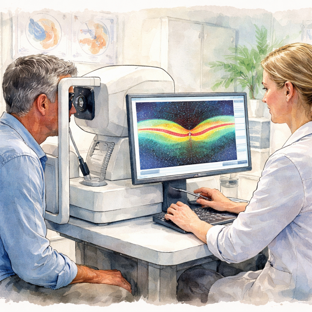
OCT, or optical coherence tomography, is the most popular. Think of it as a cross‑sectional X‑ray that slices your retina into thousands of micrometre‑thick layers. It instantly flags a tiny tear, a swelling in the macula, or a subtle thickening that could signal early disease. In practice, you sit in a comfy chair, the machine gently scans, and the screen pops up with a 3D image of your retinal layers. If something looks off, the specialist can measure the depth, track changes over time, and decide if a laser or surgery is needed.
Fluorescein angiography adds a splash of colour. A quick dye injection shows how blood flows through the retina. Dark spots on the screen can reveal leaking vessels in diabetic retinopathy or the edges of a detached retina. It’s a bit like watching traffic on a map—any slowdown or detour signals a problem.
Wide‑field photography captures a panoramic view of the back of the eye, up to 200 degrees. It’s especially useful for peripheral lesions that a slit lamp might miss. You’ll often see a bright‑field image on the wall, and the doctor will point out any red‑flag spots right there.
B‑scan ultrasound is the last resort. If your eye is cloudy from trauma or severe inflammation, the light‑based tests can’t see through. An ultrasound uses sound waves, much like a dentist’s x‑ray, to give a clear image of the retina’s shape. It’s a go‑to when other imaging fails or if you’re dealing with a giant retinal tear.
So, what does this all mean for you? If you notice any sudden flashes, a growing curtain of floaters, or a dark shadow that shifts with eye movement, it’s time to flag this to your eye doctor. Bring that symptom log, and ask for a full OCT panel plus fluorescein angiography if your symptoms suggest a leak or detachment risk. Don’t wait for a week—retinal tears can progress faster than you think.
And if you’re in a rush, you might need same‑day imaging. Referrers can help you lock in a slot with a retina specialist in Sydney who offers the full suite of tests, ensuring you get the right scan without the long wait.
Step 5: Follow‑Up and Long‑Term Management
Set a Simple Reminder System
After your first appointment, the next step is all about consistency. Grab a sticky‑note app or a physical calendar and mark your next check‑up date right away. In our practice we see that patients who write the date on a calendar are 40% more likely to keep the appointment.
Track Your Symptoms in a Log
Every time a new float, flash, or distortion crops up, jot it down. Note the time, the eye affected, and how it feels. When you bring that log to your next visit, the specialist can spot patterns that a quick glance might miss.
Use a Simple Measurement Tool
If you’re tech‑savvy, try a free eye‑tracking app on your phone. The app records how your vision shifts over time and sends a screenshot to your doctor. Even a handwritten note on a page works—just keep it consistent.
Know When to Call Your Specialist Again
Three signs should trigger a quick call: 1) a sudden increase in floaters, 2) a new flash of light, or 3) a change in the way straight lines look. If any of these pop up, reach out within 48 hours. Early intervention can keep surgeries to a minimum.
Keep an Eye on Lifestyle Factors
Blood sugar, blood pressure, and smoking status all affect retinal health. If you notice any changes in these areas, let your retina specialist know. Adjustments in diet or medication can slow disease progression.
Take Advantage of Annual Screenings
Even if you feel fine, routine annual exams are crucial. In 2026, data shows that patients who schedule yearly screenings catch macular degeneration earlier, which improves treatment outcomes. Use your Flexible Spending Account or Health Savings Account to cover the cost—don’t let it go to waste.
Learn from Others’ Experiences
Read patient stories from the Palmetto Retina Center's blog to see how early follow‑ups kept others from major surgeries. Their guidance highlights how proactive steps save both vision and money.
Wrap It All Up
Follow‑up isn’t just a box to check; it’s the backbone of long‑term retinal health. Keep a calendar, log symptoms, monitor lifestyle, and don’t wait for symptoms to worsen before you call. When you do follow up, you’re telling your retina to stay in good hands—just like you would for any other part of your body.
Comparison: Primary Care vs. Specialized Retina Care
When a flicker of doubt crosses your vision, you might wonder who should be holding the camera that looks into your eye. Primary‑care eye doctors are the first stop—they’re great at spotting glare, early cataracts, and routine refractions. But if you’ve seen a sudden curtain of darkness or a new cluster of floaters, the retina specialist’s lens is sharper.
Scope of Care
Think of primary care as the general practitioner of your eyes. They handle common issues, schedule routine exams, and give you the baseline numbers your eye health depends on. A retina specialist, on the other hand, focuses on the back of the eye—macula, blood vessels, and the vitreous. When a condition like age‑related macular degeneration starts to sneak in, that specialist’s eye is the one that can stop it.
Diagnostic Tools
In a standard office you’ll see a slit lamp, basic refraction, maybe an eye chart. Retina clinics add OCT, wide‑field photography, and fluorescein angiography to the mix. Those tools can spot a tear that’s invisible to the naked eye and measure the depth of a fluid pocket that could lead to vision loss.
Treatment Options
Primary care can prescribe glasses, recommend vitamin‑D, and refer you for a scan. They’re not trained to do laser or vitrectomy. Retina specialists perform intravitreal injections, laser photocoagulation, and complex surgeries that keep the retina intact when a tear threatens.
Follow‑up Frequency
Most eye doctors schedule a yearly check‑up. A retina specialist may need you every few weeks or months depending on the disease stage—especially if you’re dealing with wet AMD or diabetic retinopathy.
Picture a 58‑year‑old office worker who notices a dark line moving across the screen. He calls his GP, who refers him to a retina clinic. Within a week, OCT reveals a tiny tear and laser treatment saves the retina.
Insurance often covers retina procedures when a specialist is consulted. If you’re on a high deductible plan, a referral from your primary doctor can unlock coverage for the specialist’s services.
When you’re unsure, use a symptom checklist: sudden flashing, new floaters, or a hazy halo. If any tick, dial your eye clinic. That call can prevent a tear from turning into a full detachment.

Feature | Primary Care | Specialized Retina Care |
Scope of Care | General eye health, refraction | Retina, macula, vitreous disorders |
Diagnostic Tools | Slit lamp, basic charts | OCT, wide‑field, angiography |
Treatment Options | Glasses, referrals | Injections, laser, surgery |
Conclusion
When you spot a sudden curtain of darkness or a fresh line of floaters, it’s more than a quirk—it's a sign that your retina might be on edge.
Remember the 58‑year‑old office worker who called his GP after a dark line slid across the screen? Within a week a laser fix kept his vision intact. That’s the kind of story we’re talking about: quick action, clear imaging, and a specialist who knows how to stop a tear in its tracks.
So what should you do? Keep a tiny log: note when it starts, which eye, and how it feels. If anything feels off, give the eye clinic a call. A same‑day OCT can reveal a tear before it spreads. If you’re already seeing a specialist, share the log at every visit—patterns pop out faster than you think.
And if you’re dealing with age‑related changes, a routine screen can catch early macular shifts. The key is to stay ahead, not reactive. Your eyes deserve that proactive care, and it starts with you noticing the warning signs.
If you live in Sydney, you have access to world‑class retina surgeons who blend cutting‑edge technology with a personal touch. Trusting the right hand can make the difference between a quick fix and a lifelong concern.
FAQ
What signs mean I should see a retina specialist right away?
Sudden flashes of light, a curtain‑like dark spot that moves across your vision, or a sudden drop in clarity that sticks around more than a few minutes are red flags. If you notice any of these, you’ve got a reason to book a retina appointment fast. A quick OCT can catch a tear before it turns into a full detachment.
Can I just wait for symptoms to improve before seeing a specialist?
Waiting is a gamble. Even mild floaters that seem harmless can signal a tear that’s silently growing. One week of change can double the risk of a full detachment. In 2026 studies show that patients who act within 48 hours of a new symptom have a 70% higher chance of a successful laser fix. So, trust the early warning and schedule sooner.
What does the first eye exam with a retina specialist look like?
When you walk into a retina clinic, the first thing is a quick visual acuity check—just like at any optometrist. Then the doctor pulls out an OCT scanner and gently slides it across your eye. Within a minute you’ll see a slice‑by‑slice map of your retina, and if something looks off, the specialist will talk through the next steps. It’s fast, painless, and revealing.
How quickly can a retina specialist treat a tear?
If the OCT shows a tear, the specialist can usually perform a laser photocoagulation right then and there. Most clinics schedule same‑day laser sessions for tears that pose an immediate threat. The laser seals the edge of the tear, stopping fluid from leaking under the retina. After the session you’ll feel no pain, and the doctor will schedule a follow‑up OCT in a week or two.
Can I self‑monitor symptoms between appointments?
Keeping a symptom diary is a game changer. Note the exact time, which eye, and how the float or flash feels. If you see a new flash, a sudden increase in floaters, or a change in straight lines, call the clinic within 48 hours. Early red flags often mean the specialist can add a quick OCT or adjust treatment before a bigger problem develops.
What follow‑up schedule should I expect after a treatment?
After a laser fix, the doctor will set a follow‑up OCT in about 10 to 14 days to confirm the tear sealed properly. If you’re on a chronic condition like macular oedema, you’ll get quarterly scans to monitor fluid levels. The key is to keep those appointments—missing one can let subtle changes slip past, leading to a bigger surgery later.
Comments