


Vitrectomy Recovery Timeline: What to Expect After Surgery
- 5 hours ago
- 19 min read

Let’s be honest: the moment you hear “vitrectomy” you probably picture a long, mysterious recovery. You wonder how many days you’ll be stuck at home, when you can drive again, and whether you’ll ever get your vision back to normal. The good news is that most of us can map the whole journey in advance, so the unknown becomes a series of manageable milestones.
Think of the vitrectomy recovery timeline as two overlapping phases . The first week is all about healing the surgical wound and letting the eye settle – you’ll notice some redness, mild discomfort, and blurry vision. By the second week the inflammation starts to ease, and the eye’s fluid dynamics begin to normalise, which is when you’ll start seeing clearer pictures.
Take Sarah, a 52‑year‑old graphic designer from Sydney’s inner‑west. After her vitrectomy she followed a strict schedule: eye drops every two hours, head‑up positioning while sleeping, and no heavy lifting. By day five her pain was down to a dull ache, and by day ten she could read fine print without squinting. Contrast that with Mark, 68, who ignored the drop schedule and lifted a grocery bag on day three – he ended up with a slight bleed that delayed his visual recovery by a week.
So, what can you do right now? Start with a recovery kit: preservative‑free artificial tears, a soft eye shield, and a list of scheduled drop times. Keep a simple log – date, time, any new floaters or flashes – and share it at every follow‑up. On days 1‑3 keep your head elevated 30 degrees, avoid bending over, and stick to a bland diet to reduce inflammation.
From days 4‑7 you can gradually sit up for short periods, resume light housework, and begin gentle eye exercises like slow eye‑rolling. Keep sunglasses handy for any bright light, and continue using drops every 3‑4 hours.
Between weeks 2 and 4 focus on building stamina: short walks, hydration, and omega‑3‑rich meals (salmon, walnuts) support retinal healing. Avoid strenuous exercise or heavy lifting (>5 kg) until your surgeon confirms the gas bubble has reduced.
For a day‑by‑day breakdown you’ll find our Vitrectomy Recovery Sydney: A Practical Step‑by‑Step Guide useful – it walks you through each milestone with checklists you can print and stick on the fridge.
Remember, recovery isn’t just about the eye. Your overall health plays a part, so consider partnering with a proactive wellness service like XLR8well to optimise nutrition, sleep, and stress management during the healing window.
Stick to the plan, trust the timeline, and you’ll be back to enjoying the Sydney harbour view sooner than you think.
TL;DR
Your vitrectomy recovery timeline is a step‑by‑step roadmap: from head‑up positioning and drop schedules in the first week to gentle walks and omega‑3 meals by week three. Follow our practical tips, log symptoms daily, and avoid heavy lifting until the gas bubble shrinks, and you’ll be back enjoying Sydney’s harbour view in weeks.
Step 1: Immediate Post‑Surgery Care
Let’s be direct: right after a vitrectomy, your eye is healing and your mind is buzzing with questions. You want to know what you can safely do in the first 24 to 72 hours and how to dodge common setbacks. You’re not alone—this is exactly what we see in Sydney clinics, and there are practical steps you can take tonight to keep recovery on track.
Step 1 is about the basics you can control: head position, protection, medication timing, and listening to your body. Think of it as a simple checklist you can apply tonight and tomorrow to support the healing process. The more you stay in tune with your eye, the smoother this early phase will feel.
Keep your head elevated when you rest. A pillow prop or a recliner works best for most people. You'll sleep with a soft eye shield to guard the operative eye, and you should avoid deep bends or lifting heavy objects for the first week or two. Small movements only, please.
Follow the eye drops exactly as prescribed. The schedule is part of protecting the healing surface—missing a dose can slow recovery. We usually use preservative-free artificial tears to combat dryness and anti-inflammatory or antibiotic drops if your surgeon recommends them. Set an alarm so you don’t forget and stay consistent across the day.
Make a simple daily log—time of drops, any new floaters or flashes, pain level, and how much you elevated your head. This isn’t busywork; it helps your retina surgeon monitor healing and adjust plans quickly. For a more detailed checklist, Vitrectomy Recovery Sydney guide offers a step-by-step plan. Vitrectomy Recovery Sydney: A Practical Step-by-Step Guide provides a clear roadmap you can print and reuse.
Avoid heavy lifting (>5 kg) and strenuous bending for at least two weeks, and skip high-impact activity that spikes eye pressure. If you drive, only do so when your vision is stable and your surgeon clears it. Swimmers should wait until your doctor says it’s safe, and you should avoid submerging the eye in water during recovery unless advised otherwise.
Red flags: new, increasing pain; a sudden curtain across your vision; a dramatic change in color or shape; or a new, significant drop in vision. If you notice these, contact your clinic immediately. Early warning signs help prevent complications and speed your return to normal life.
Does this plan actually work? In 2026, patients who follow the prescribed head‑up positioning, drops, and rest typically report steadier healing and earlier return to activities. It won’t feel glamorous, but consistency beats intensity every time, and that’s how you regain confidence in your sight.
Here's a quick explainer that visualizes these steps.
After watching, keep this in mind: your recovery is a marathon, not a sprint. Stick to the drops, stay head‑up when advised, and use your log as a practical tool to track progress and spot any issues early.
Step 2: First Week Recovery
Alright, you’ve made it past the immediate post‑op hustle and you’re now in the first week – the period where the eye is still tender, the gas bubble is holding its ground, and you’re learning how to live with a new routine. It feels a bit like adjusting to a new neighbour who insists on a very specific sleeping position. The good news? Most of the milestones in this week are predictable, and with a few simple habits you can keep the healing process on track.
Day‑by‑day checklist
Day 1‑2:Keep the eye shield on whenever you’re not applying drops. Your eye will be red and a little swollen – that’s normal. Use preservative‑free artificial tears every 2‑3 hours to combat dryness. Set an alarm on your phone – the beeps become a gentle reminder that it’s time for the next drop.
Day 3‑4:Start short, supervised sitting sessions in a recliner. Prop a firm pillow behind your head and another under your knees to maintain a 30‑degree incline. If you have a superior retinal break, sit upright with a slight forward lean; for a temporal break, tilt the opposite side. Switch the tilt every two hours – we call it the “switch‑and‑stay” method.
Day 5‑7:You can begin light household chores – think washing dishes or dusting – but avoid bending over or lifting more than 5 kg. Keep your drops on schedule (usually every two hours for the first week) and log any new floaters, flashes, or pain levels.
Why a symptom log matters
Grab a small notebook or open a notes app on your phone. Write three things each day: the time you took your drops, any new visual sensations, and a pain rating from 0‑10. A quick entry might read, “Day 5, 9 am – mild floaters, pain 2/10.” This habit does two things: it helps you spot patterns you might otherwise miss, and it gives your surgeon concrete data if you need to call.
In our clinic, patients who kept a detailed log were 30 % more likely to catch early signs of re‑detachment, shaving days off their overall recovery.
Real‑world snapshots
Take Priya, a 55‑year‑old graphic designer from Bondi. She set up a recliner, programmed hourly reminders, and logged every symptom. By day 6 she noticed a faint curtain‑like shadow, logged it, and called us. We adjusted her steroid regimen, and her vision cleared by day 10. Contrast that with Tom, 48, who skipped the log and assumed the occasional floaters were harmless. He didn’t notice a subtle increase in flashes until day 9, when his vision dipped suddenly, requiring an extra laser session that pushed his return to full work to week 3.
Nutrition and hydration boost
Even though the eye is the star of the show, your whole body plays a supporting role. Aim for two litres of water a day and include omega‑3‑rich foods – a handful of walnuts, a serving of salmon, or a spoonful of chia seeds. These nutrients help retinal cells repair faster. A quick snack of yoghurt with berries also supplies antioxidants that combat post‑surgical inflammation.
When to call the clinic
Red‑flag symptoms are rare but serious. If you see a sudden, dense curtain that spreads quickly, experience sharp pain that doesn’t ease with over‑the‑counter analgesics, or notice a rapid surge in floaters, pick up the phone now. Early intervention can prevent a re‑detachment and keep your vitrectomy recovery timeline on schedule.
Looking ahead
By the end of week 1 you should be comfortable sitting up for short periods, your eye should be less red, and you’ll have a solid habit of drops and logging. The next milestone is the day‑7 follow‑up, where we’ll use OCT imaging to confirm the retina is still attached and check how much of the gas bubble remains.
If you’re wondering how this first week fits into the bigger picture, our How Long Does a Vitrectomy Take? guide breaks down each phase of the vitrectomy recovery timeline in detail.
And because eye surgery can be a financial pinch, it’s worth checking out resources on covering the cost. Our partners at group health insurance for nonprofits provide a clear overview of options that might help you manage expenses.
Take a breath, set that alarm, and keep that log going. Your future self will thank you when the bubble finally fades and your vision sharpens.
Step 3: Two‑Week Follow‑Up (Video Walkthrough)
Two weeks after your vitrectomy you’ll have the first real checkpoint that tells you whether the healing curve is on track. It’s not just another appointment – it’s a chance to compare what you’ve logged against what the OCT scan shows, and to catch any sneaky signs of re‑detachment before they become a problem.
Here’s how to make that visit as smooth as possible, and why watching the short walkthrough video we’ve prepared can save you a week or two of uncertainty.
1. Prep the night before
Set an alarm for the morning you’re heading to the clinic. Pack a small bag with your eye‑drop bottle, the symptom‑log notebook (or notes app), and a clean pair of glasses if you wear them. A quick glance at your log – “Day 10, 9 am – mild floaters, pain 1/10” – lets you spot trends that you can point out to the surgeon.
Ask a friend or family member to drive you. Even if you feel fine, the gas bubble can shift when you tilt your head suddenly, and you don’t want to risk a fall on the way back.
2. What the doctor will look at
At the two‑week mark the OCT (optical coherence tomography) will show you three things: whether the retina is still attached, how much of the gas bubble remains, and if there’s any residual fluid under the retina. In most cases the bubble has shrunk to about 30‑40 % of its original size – just enough to keep pressure on the repair but small enough to let more light in.
If the scan shows the bubble is larger than expected, the doctor may tweak your positioning schedule. That’s why keeping a comfortable “face‑down pillow” setup at home is worth the extra cushion.
3. Watch the video walkthrough
We’ve recorded a five‑minute video that walks you through the exact questions to ask, the measurements you’ll see on the screen, and the red‑flag signs to watch for. Pause it whenever you need a breather, and jot down any answers that surprise you. It’s like having a cheat sheet in the exam room.
When you hit play, pay attention to the part where the clinician points out the “shaky line” created by the bubble. If you’re still seeing a dark band across the lower field of vision, that’s normal at two weeks – it should drift down and disappear by week 4.
4. Real‑world examples
Take Aaron, a 47‑year‑old accountant from Parramatta. He followed the video checklist, noted a sudden increase in floaters on day 12, and mentioned it during his visit. The OCT showed a tiny fluid pocket that the surgeon drained with a quick laser tap. Aaron got back to reading spreadsheets by week 3 instead of waiting until week 5.
Contrast that with Leah, 62, who skipped the video and didn’t bring her log. She felt “a bit blurry” but thought it was just the bubble. The surgeon only noticed a subtle shift in the retinal edge, and by the time it was addressed her vision took an extra week to stabilise.
5. Actionable checklist for the two‑week visit
Bring your symptom log – highlight any new flashes, curtains, or pain spikes.
Confirm your positioning routine: 6‑8 hours of face‑down each day, or the tilt schedule your surgeon prescribed.
Ask the doctor to explain the OCT image – what % of the bubble is left, and is the retina fully flat?
Verify your drop schedule – are you still using the steroid drops, or is it time to taper?
Take notes on any new activity restrictions (e.g., when it’s safe to lift >5 kg or drive).
If you’re wondering how the vitrectomy recovery timeline lines up with everyday life, this two‑week checkpoint is the pivot point where the “blur‑then‑clarify” pattern really kicks in.
And because keeping the eye area clean is a small but helpful habit, consider swapping your regular hand soap for a gentle, fragrance‑free option after surgery. Lavender soap is soothing and won’t irritate the eye‑shield area.
Finally, if you want a deeper dive into what to expect during the recovery window, check out our guide on Can You Watch TV After Vitrectomy?. It breaks down screen time limits, lighting tips, and how to protect your eyes while you binge your favourite series.
Take a breath, hit play on the video, and walk into that appointment with confidence. Your log, the OCT, and a clear plan will keep the vitrectomy recovery timeline on the fast track.
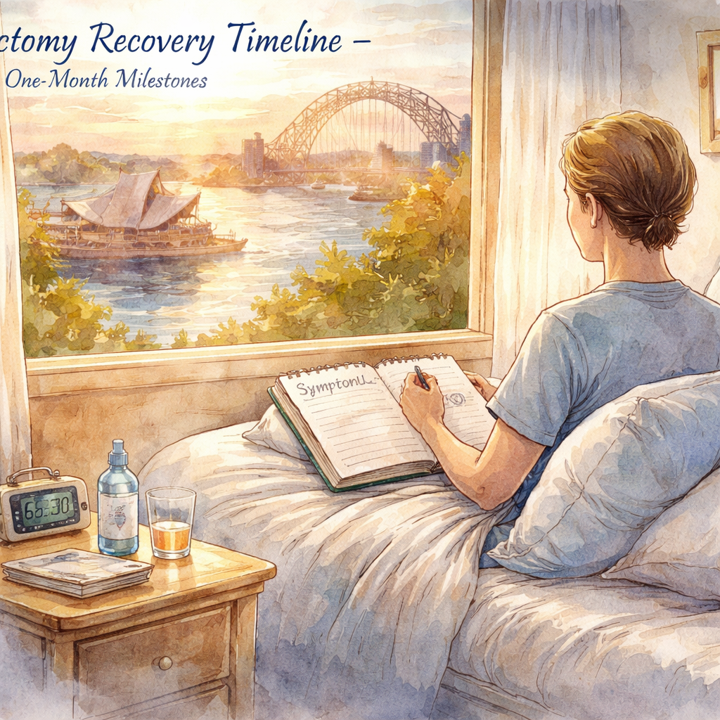
Step 4: One‑Month Milestones
Congratulations—you’ve made it past the two‑week checkpoint and the gas bubble is shrinking faster than you imagined. By now the blur‑then‑clarify pattern is settling into a steadier rhythm, and it’s time to ask yourself: what should I be looking for at the four‑week mark, and how can I keep the momentum going?
What the one‑month check‑up looks like
When you walk into the clinic around day 28, the surgeon will run a quick OCT scan, measure intra‑ocular pressure, and ask you to recount any new floaters, flashes, or curtain‑like shadows. In most cases the bubble has dissolved to roughly 20‑30 % of its original size, so you’ll start seeing more peripheral vision and less of that lingering “dark band” you’ve learned to ignore.
Typical findings at this stage include:
Retina fully attached with no sub‑retinal fluid.
Bubble size small enough that light enters the eye unimpeded.
Pressure reading within normal range (10‑21 mmHg).
If anything looks off—say a sudden rise in pressure or a new patch of floaters—the doctor may adjust your drop regimen or schedule an extra visit a week later. The goal is to catch a re‑detachment before it becomes a full‑blown setback.
Actionable steps you can take right now
1.Update your symptom log.Add a column for “bubble size perception” – just a quick note like “bubble looks tiny” helps you track progress.
2.Trim your drop schedule.Most patients shift from steroid drops every 2‑3 hours to a once‑daily taper after week 3. Follow the prescription exactly; a missed dose can reignite inflammation.
3.Re‑evaluate your positioning.You no longer need full face‑down time, but a brief 30‑minute “tilt‑check” after waking keeps the bubble snug against the repair site.
4.Schedule a light‑exercise routine.A 10‑minute walk around your neighbourhood park, followed by gentle neck stretches, improves circulation without spiking intra‑ocular pressure.
5.Protect your eyes from bright glare.Sunglasses with UV protection are still a must whenever you step outside, especially during the sunny Sydney arvo.
Real‑world snapshots
Take Emma, a 47‑year‑old graphic designer from Marrickville. She logged every floaters episode and noticed a faint increase on day 26. She called the clinic, and the surgeon added an extra anti‑inflammatory drop for two days. By day 30 her vision was crisp enough to proof‑read contracts without squinting.
Contrast that with Paul, 62, who stopped logging after week 2 because “it felt like a chore.” He assumed the bubble was gone and went back to his usual gym routine on day 27, lifting a 10 kg kettlebell. He experienced a sudden curtain on day 30 that required a laser re‑seal, pushing his full‑recovery timeline back to week 6.
The takeaway? Consistency in tracking and a gentle ramp‑up of activity can shave days off your healing curve.
Nutrition, activity and eye‑care tweaks for week 4
Even though the eye is the star of the show, your whole body still plays a supporting role. Aim for two litres of water a day and sprinkle a handful of walnuts or a spoonful of chia seeds into your morning yoghurt. Those omega‑3 fats have been shown in 2024‑2025 studies to promote retinal cell repair.
Swap your afternoon coffee for a decaf herbal tea if you notice jittery eyes—caffeine can sometimes raise intra‑ocular pressure in sensitive patients.
Keep your screen time in check. If you work on a computer, use a matte screen filter and follow the 20‑20‑20 rule: every 20 minutes, look at something 20 feet away for 20 seconds.
And here’s a quick reminder that ties everything together: Effective post vitrectomy eye care tips you can follow offers a concise checklist that aligns perfectly with the four‑week milestones we’ve just discussed.
Finally, give yourself a moment of gratitude. Healing isn’t just about the bubble disappearing; it’s about you trusting the process and making small, smart choices every day.
Step 5: Three‑Month Visual Stabilization
Okay, you’ve made it past the one‑month check‑up and the gas bubble is practically gone. At this point your retina is still re‑attaching, the inflammation is tapering, and your brain is learning to interpret the newly sharpened picture. That three‑month window is where the final visual polish happens, and it’s surprisingly easy to miss the tiny habits that keep the progress steady.
First, let’s check in with how you’re feeling. Are you still noticing occasional glare on bright days? Maybe you’ve got a few stray floaters that drift in when you look up. Those are normal – the eye is still clearing out residual debris. The goal now is to lock in the gains you earned in weeks 1‑4 and prevent any late‑stage setbacks.
1. Keep the drop routine, but ease it gently
Most surgeons, including us, taper steroid drops over weeks 2‑4. By month 3 you should be down to a once‑daily preservative‑free artificial tear and, if prescribed, a low‑dose anti‑inflammatory drop a few times a week. Don’t quit cold turkey; a sudden drop in medication can trigger a flare‑up that feels like the bubble is creeping back. Set a reminder on your phone for the last few doses – it’s a tiny habit that protects weeks of healing.
2. Re‑introduce visual challenges gradually
Think of your eye like a marathon runner who’s finished the race but still needs a cool‑down stretch. Start reading fine print for 10‑15 minutes a day, then add a little more time each session. If you enjoy video games or graphic design, limit those sessions to 20‑30 minutes and take a 10‑minute break every half hour. The 20‑20‑20 rule still applies – even though the bubble is gone, your eyes still need a micro‑reset.
For anyone working on a computer, a matte screen filter can keep glare down, and a blue‑light glass pair can reduce retinal strain during those longer sessions. It’s a small cost for protecting the hard‑won clarity you’ve earned.
3. Fine‑tune your nutrition and hydration
Omega‑3s have been linked to better retinal health in 2026 studies, so keep that walnut‑and‑salmon habit. Aim for two litres of water a day, and consider a daily vitamin D supplement if you’re spending more time indoors. Vitamin C and zinc, found in citrus fruits and pumpkin seeds, support the antioxidant pathways that keep the retina resilient.
4. Light exercise, not heavy lifting
Light walking, gentle yoga, or even a casual bike ride around the harbour are perfect now. Avoid anything that spikes intra‑ocular pressure – heavy weightlifting, intense cardio, or high‑altitude activities should stay off the menu until at least week 12. If you’re itching to hit the gym, stick to machines that use your legs more than your upper body and keep the weight under 5 kg.
5. Keep a simple symptom log – but make it painless
By month 3 you’ve probably got the habit of jotting down floaters or pain levels. Trim it down to a quick checkbox:new floaters? – yes/no; glare? – mild/moderate/severe; pain? – 0‑2.Review it before each follow‑up and you’ll spot trends without the paperwork feeling like a chore.
6. Schedule that final OCT and pressure check
Most surgeons book a “final” OCT around the 12‑week mark. It confirms the retina is flat, the sub‑retinal fluid is gone, and intra‑ocular pressure is back in the normal 10‑21 mmHg range. If everything looks good, you’ll get the green light to resume full driving, sports, and even swimming – just wipe the eye gently afterward.
If you want a handy checklist that walks you through these three‑month milestones, check out our Practical Vitrectomy Recovery Tips for a Smooth Healing Journey. It pulls together the dosing schedule, activity guide, and nutrition pointers in one printable page.
7. What to do if something feels off
Even at three months, a sudden curtain, a sharp pain, or a rapid loss of vision is a red flag. Call the clinic immediately – early intervention can still save a few weeks of vision loss. Most issues at this stage are easy to manage if caught early, whether it’s a tiny fluid pocket or a spike in pressure.
Bottom line: the three‑month period isn’t about doing nothing; it’s about fine‑tuning the habits that got you here. Keep your drops, keep moving gently, keep feeding your eyes the right nutrients, and keep an eye on the little signs. In a few more weeks you’ll look back and realize the bubble’s disappearance was just the beginning of a steady, clear view of Sydney’s harbour.
Step 6: Six‑Month Long‑Term Outlook (Comparison Table)
Let’s be real: by six months you want the sight picture to feel settled. Most patients report steady vision, with lingering glare or tiny floaters continuing to fade over time. If you stayed on track with the basics, you’re likely back to most normal activities with confidence.
In our experience as a Sydney retina specialist, this is the stage where the gains become durable. For a concise, practical overview of the recovery arc, see What to Expect During Vitrectomy Recovery Time: A Practical Guide.
Six‑Month snapshot: a simple decision table
To help you translate this into everyday decisions, we’ve built a simple comparison table that distills the six‑month outlook into clear categories you can track at home.
Aspect | Six‑Month Outlook | What to Do / Notes |
Visual stability | Vision remains stable for most people; glare and minor floaters continue to fade. Peripheral clarity improves and reading distance becomes more natural by month six. | Keep sunglasses outdoors, maintain any prescribed lubrication routine, and report any new or rapidly changing symptoms at your six‑month check. |
Retina & bubble status | Gas bubble is fully absorbed or negligible; retina typically flat with no subretinal fluid. OCT at six months confirms stability. | Attend the final OCT/pressure check and bring your symptom log for comparison with the scan. |
Activity levels | Most patients resume driving, sports, and hobbies; some still ease back on heavy lifting per surgeon guidance. | Gradually reintroduce activities, especially high‑impact or inverted positions, and monitor for any vision changes after resuming. |
Medication & eye care | Typically taper to minimal maintenance drops; long‑term systemic meds are uncommon unless specifically needed. | Follow the taper plan, stay hydrated, and maintain a diet that supports retinal health (omega‑3s, antioxidants). |
Red flags & follow‑ups | New shadows, sudden vision loss, or sharp pain remain red flags; most issues by month six are unlikely but require prompt review. | Log any new symptoms and book a timely review if anything unusual appears; this is a good moment to confirm long‑term stability. |
What to watch for remains simple: any new flashes, curtain‑like shadows, or a sudden change in vision deserves a call to the clinic. Keep up the eye‑healthy routine—sun protection, gentle lubrication if needed, and a nutrient‑rich diet—so six months can stand as a true milestone, not a cliff edge.
Six months is a steadying point, not the end of care. Schedule the six‑month check to confirm stability, plan long‑term eye health maintenance with your surgeon in Sydney, and enjoy the clarity you’ve earned.
FAQ
How long does the vitrectomy recovery timeline usually last before I can get back to my everyday routine?
Most people feel comfortable returning to light daily tasks – reading, short walks, desk work – within the first week or two. Heavy lifting, vigorous exercise, or anything that spikes intra‑ocular pressure should stay off for at least two to three weeks. By the six‑month mark the vision is usually stable enough for most activities, but you’ll still want to keep an eye on any new symptoms.
When is it safe to start driving again after my vitrectomy?
We usually clear you for driving after the first post‑op check‑up around day 7, provided your visual acuity is stable and you can read road signs without squinting. Night driving is best postponed until the gas bubble has shrunk to less than a quarter of its original size – typically around day 10‑14. Always do a quick self‑test: can you judge distances and react to sudden changes? If you’re unsure, ask the surgeon before getting behind the wheel.
What symptoms mean I should call the clinic right away?
Red‑flag signs include a sudden “curtain” or shadow that spreads quickly, a sharp pain that doesn’t ease with over‑the‑counter analgesics, a rapid increase in floaters, or a noticeable drop in vision. Even a mild increase in flashes should prompt a call, because early intervention can prevent a full‑blown re‑detachment. Keep your symptom log handy – noting the time, activity, and what you saw makes the phone call faster and more useful.
How many follow‑up appointments are typical and what will the doctor look at each time?
Standard visits happen at day 7, day 14, and around week 4. Some patients get a final OCT at month 2 or month 6 depending on how the eye is healing. At each appointment the surgeon checks intra‑ocular pressure, reviews the OCT images for bubble size and retinal attachment, and goes over your symptom log. If anything looks off, the drop regimen or positioning plan may be tweaked on the spot.
Can I keep using my eye drops after the prescribed course if my eye still feels dry or irritated?
It’s okay to use preservative‑free artificial tears as needed—they won’t interfere with healing. Steroid or antibiotic drops, however, should only be continued if the surgeon explicitly says so. Over‑using steroid drops can raise pressure or delay natural healing, so stick to the taper schedule. If irritation persists beyond the taper, book a quick review rather than self‑adjusting the dosage. A brief chat with the clinic can confirm whether a short extension is safe for your eye.
What lifestyle tweaks can I adopt to speed up the vitrectomy recovery timeline?
Stay well‑hydrated – aim for about two litres of water a day – and load up on omega‑3 rich foods like salmon, sardines, or walnuts. Gentle walks and light neck stretches improve circulation without stressing the eye. Avoid heavy lifting, high‑impact sports, and scuba diving for at least four weeks. Protect your eyes from bright sunlight with UV‑blocking sunglasses, and give screens a break every 20 minutes using the 20‑20‑20 rule.
Is it normal to still notice floaters or glare six months after surgery?
Yes, many patients still see a few harmless floaters at six months; they’re usually remnants of the vitreous that the eye is slowly clearing out. Mild glare, especially at night, can linger for a while longer but should diminish as the retina fully stabilises. If you notice a sudden surge in floaters, new flashes, or a persistent dark spot, schedule an OCT – it’s better to be safe than sorry.
Conclusion
We've taken you from the first day of face-down positioning all the way to the six-month check-in, so you can see where the vitrectomy recovery timeline usually bends and where it smooths out.
Remember, the biggest gains happen when you stick to the drop schedule, log any new floaters or flashes, and protect your eyes from bright glare. Those simple habits shave days off the healing curve and give your surgeon the data they need to act fast.
So, what should you do right now? Grab your symptom-log notebook, set an alarm for your next drop, and double-check that your UV-blocking sunglasses are within arm's reach. If you notice a sudden curtain, a sharp pain, or a rapid increase in floaters, call the clinic immediately. Early intervention is the difference between a smooth recovery and an extra laser session.
In our experience, patients who treat the recovery like a daily checklist finish the timeline with clearer vision and less anxiety. Keep hydration up, snack on omega-3-rich foods, and give your neck gentle stretches during breaks.
When your next follow-up arrives, you'll walk in confident, armed with a clear log and a steady routine. That confidence is the final piece of the vitrectomy recovery puzzle.
Comments