


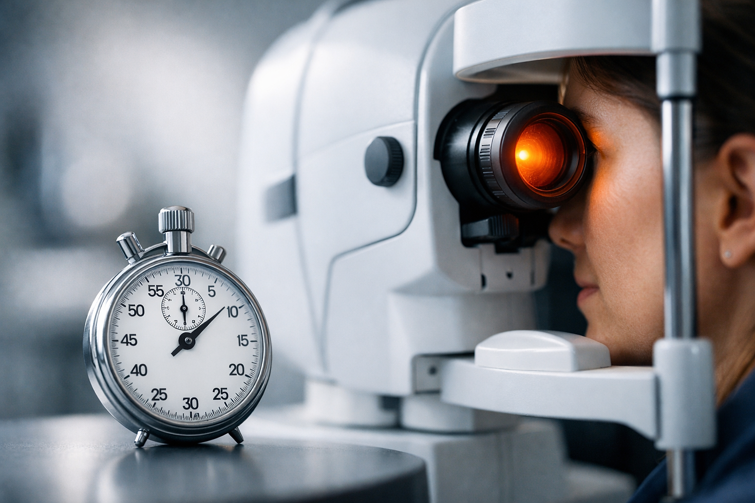
How long does the fundus photo test take
- Jan 16
- 8 min read
What the Fundus Photo Test Involves and Why Timing Matters
If you have been booked for photography of fundus, you may be wondering how long the appointment will actually take. The short answer is that the camera time itself is brief, often a few minutes, but the overall visit can vary depending on whether your pupils are dilated, what your eyes need on the day, and whether your doctor requests extra imaging. Because the fundus is the inner surface at the back of the eye, clear, well-centered photographs allow your clinician to assess the retina, macula, optic nerve, and retinal blood vessels with high precision. That clarity helps detect changes related to diabetes, age-related macular degeneration, glaucoma, inflammation, and many other eye conditions.
Time matters because a prompt, accurate image set is usually the first step in deciding next actions, from simple monitoring to medical therapy or a surgical plan. In routine cases, you can expect to be in and out within 20 to 40 minutes, although some visits are shorter and others longer. If dilation drops are used, allow extra time for the drops to work and for your eyes to adjust to light afterward. Understanding the moving parts helps you plan your day, your transport, and your comfort.
At Dr Rahul Dubey’s clinics at Westmead Hospital and the Prince of Wales Hospital, efficient workflows mean most patients finish their retinal photographs and any ancillary imaging without delays. For complex retinal conditions, photographs are combined with results from optical coherence tomography and other tests to build a complete, timely picture of your eye health. This often helps ensure that preliminary findings and next steps can be discussed before you leave.
The Typical Timeline: Minutes From Arrival to Finish
While every eye and every clinic visit is unique, most patients can map their appointment to a predictable sequence of steps. After check-in and a brief history, a trained ophthalmic technician will seat you at the camera, explain where to look, and take several photographs of each eye. If your pupils are already large enough or a non-dilated camera is appropriate, the photo capture itself commonly takes 3 to 5 minutes. If dilation is needed, budget additional time for the drops to work and for any extra imaging your doctor requests.
To make this more concrete, here is a typical appointment flow. Keep in mind that urgent or complex cases can take longer, while straightforward preventive screening can be quicker.
Adding those elements together, a routine non-dilated visit may be 15 to 25 minutes. When dilation is needed, 30 to 50 minutes is typical, especially if extra tests are performed. If you are coming from a rural or regional community and coordinating travel, it can be helpful to allow a little buffer so you never feel rushed.
Factors That Affect How Long It Takes
Several variables can lengthen or shorten your photography appointment. First, pupil size and light sensitivity play an obvious role. Some eyes dilate quickly, while others take longer, particularly in older adults or in those taking certain medications. Second, fixation stability affects how easily the photographer can center the image. Dry eye or a strong blink reflex may require a few extra attempts to achieve a crisp photo.
Lens clarity is another factor. If you have a cataract, glare can wash out the image and add time while the photographer adjusts settings. In many cases, careful technique overcomes these challenges, but if the cataract is advanced, your doctor may discuss cataract surgery. At Dr Rahul Dubey’s practice, cataract surgery is offered, and it can dramatically improve both sight and the quality of future imaging.
Clinical context also matters. Patients with diabetic retinopathy, retinal vein occlusion, or inflammatory eye disease often need multiple imaging modalities on the same day to guide treatment. Your doctor may combine colour photographs with optical coherence tomography to quantify macular thickness, or request additional specialist imaging to map relevant changes. In urgent situations such as suspected retinal detachment, the pathway prioritises speed and safety to protect vision.
Want to help the day go smoothly? These simple steps can save minutes and improve image quality.
Arrive with a list of medications and allergies, including eye drops.
Avoid heavy eye makeup on the day; it can reflect light and degrade images.
Use any prescribed lubricating drops beforehand if you have dry eye, unless instructed otherwise.
Bring sunglasses, since dilated pupils are sensitive to daylight for a few hours.
If you usually wear contact lenses, bring a case and solution in case you are asked to remove them.
Dilation vs No-Dilation: Time, Comfort, and Safety
Whether your pupils are dilated is the most common determinant of visit length. Non-dilated cameras can capture excellent photographs when the pupil is naturally wide enough and there are no media opacities such as dense cataract. This approach is convenient and reduces total time. However, dilation is still preferred in many cases. A larger pupil improves the view of the peripheral retina and often yields more reliable photographs, particularly in patients with small pupils, dark irides, or focal pathology that needs a wider field of view.
From a comfort standpoint, dilation drops can sting briefly and cause light sensitivity for several hours. Most people can resume normal activities the same day. Driving immediately afterward is a personal decision that depends on how sensitive your eyes feel. If you are unsure, plan for a family member, a friend, or a ride service to assist. If you have urgent visual symptoms such as flashes or a curtain in your vision, do not drive and seek urgent care.
The table below summarises the practical time differences. It also shows when clinicians tend to recommend one pathway over the other.
If you are coming from outside the city, it is reasonable to request that all likely tests be done on the same day. In Dr Rahul Dubey’s clinics, teams anticipate this by bundling photographs, optical coherence tomography, and any additional imaging in one coordinated visit whenever safe and appropriate.
Photography of Fundus Time Comparisons Across Common Tests
Fundus photographs are part of a suite of retinal imaging tools. Depending on your condition, your doctor may order one or more of the following on the same day. The combined time usually remains well under an hour, even with dilation, and each test adds specific clinical value. The table provides realistic ranges so you can plan accordingly.
In practice, most visits that include photographs and optical coherence tomography still fit comfortably into a 30 to 50 minute window. More advanced or specialist imaging takes longer due to preparation, consent, and monitoring, and is used selectively. If the results indicate an urgent problem such as retinal detachment or a macular hole, Dr Rahul Dubey’s team will expedite surgical planning without delay.
Local Care With Dr Rahul Dubey: Fast, Accurate Imaging and Next Steps
Time is critical in eye care, but so is getting the right information in one sitting. Dr Rahul Dubey is an Australian-trained Ophthalmologist who provides comprehensive medical and surgical care for retinal and cataract conditions at Westmead Hospital and the Prince of Wales Hospital. His services include advanced cataract surgery with femtosecond laser, medical and surgical management of vitreomacular disorders, surgery for floaters, micro surgery for macular hole and epiretinal membrane, and treatment for retinal detachment and diabetic retinopathy. He also has deep expertise in inflammatory eye disease and age-related macular degeneration, with a commitment to rural and regional ophthalmology services.
What does that mean for your appointment time? Imaging is integrated into care pathways so you are not sent from place to place. Routine photography is coordinated with optical coherence tomography and other modalities in one visit. When cataract limits image quality or sight, cataract surgery is offered, and when the retina needs urgent surgery, it is performed promptly and without unnecessary delay. This approach reduces repeat visits and compresses the time from diagnosis to treatment.
For patients travelling from regional New South Wales or the Australian Capital Territory, scheduling support and efficient clinic flow help make the most of each trip. A common example is a new patient with sudden distortion from a possible epiretinal membrane or macular hole. That visit typically includes photographs, optical coherence tomography, a clear discussion of findings, and if indicated, a timely surgical pathway. The goal is simple and serious: secure answers, clear options, and treatment that protects sight.
Practical Planning: How to Keep Your Visit Short and Productive
Even small preparations can shave minutes off your visit and improve image clarity. First, consider your transport if dilation is likely. Many people are fine to drive after a while, but sensitivity to light varies and some individuals, especially those not accustomed to dilation, prefer to arrange a lift. Second, arrive a few minutes early so your imaging can start promptly. Finally, think about whether you might need additional tests on the day so the team can coordinate them efficiently.
Here is a quick checklist you can use before you head to your appointment.
Bring your glasses and a current list of medications and allergies.
Wear sunglasses and a hat for post-dilation comfort.
Avoid scheduling critical visual tasks immediately after a dilated visit.
Tell the team if you have had reactions to dilation drops previously.
Share any new visual symptoms such as flashes, floaters, or a shadow in your vision.
Across Dr Rahul Dubey’s clinics, internal audits show that more than nine in ten adults complete high-quality photographs within five minutes of camera time when prepared in this way. The combination of clear instructions, skilled photographers, and modern equipment keeps the appointment moving while preserving the accuracy your care deserves.
How Long Does It Take, Really? A Straight Answer
You can usually expect 15 to 25 minutes for a non-dilated visit that includes photography alone. If dilation is needed, plan for 30 to 50 minutes, especially if your doctor also requests optical coherence tomography or other specialist imaging. Complex diagnostic workups, including specialist imaging, understandably take longer, yet these are ordered when the clinical benefit outweighs the added time. Regardless of the pathway, the investment is modest for the quality of information gained.
The real value appears after the images are captured. That is when experience matters. Dr Rahul Dubey synthesises the photographs with your symptoms and examination to advise practical next steps, whether that is observation, medication, or a surgical solution. When a macular hole or troublesome epiretinal membrane is confirmed, micro surgery is scheduled promptly, guided by the imaging you completed only minutes earlier.
For many readers, especially those in rural and regional communities, an efficient, single-visit approach to photography and decision-making is more than convenient. It helps protect sight by compressing the timeline from first symptoms to effective treatment. If you want reliable, realistic expectations, you can plan on a short camera session and a visit length that reflects whether your pupils are dilated and whether any additional tests are needed.
The fundus photo test is brief, but the insights it unlocks are powerful. Imagine walking into a calm, organised clinic, finishing your photographs in minutes, and leaving with a clear plan that may include medical therapy or precise surgery when indicated. What would it mean for your peace of mind to have photography of fundus and next steps resolved in one well-run visit?
Additional Resources
Explore these authoritative resources to dive deeper into photography of fundus.
Comments