Detailed retinal assessment
Examination and imaging establish whether surgery is likely to offer a meaningful anatomical or visual benefit.
Small-incision surgery for selected conditions affecting the vitreous, retina and macula.
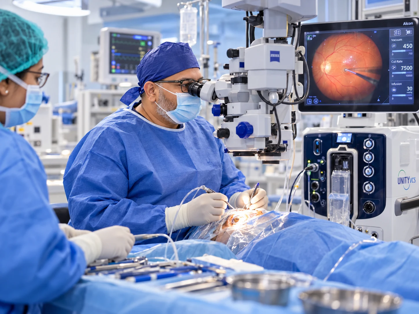
Vitrectomy removes the vitreous gel to provide access to the retina. It may be recommended for retinal detachment, vitreous haemorrhage, macular hole, epiretinal membrane and other selected conditions.
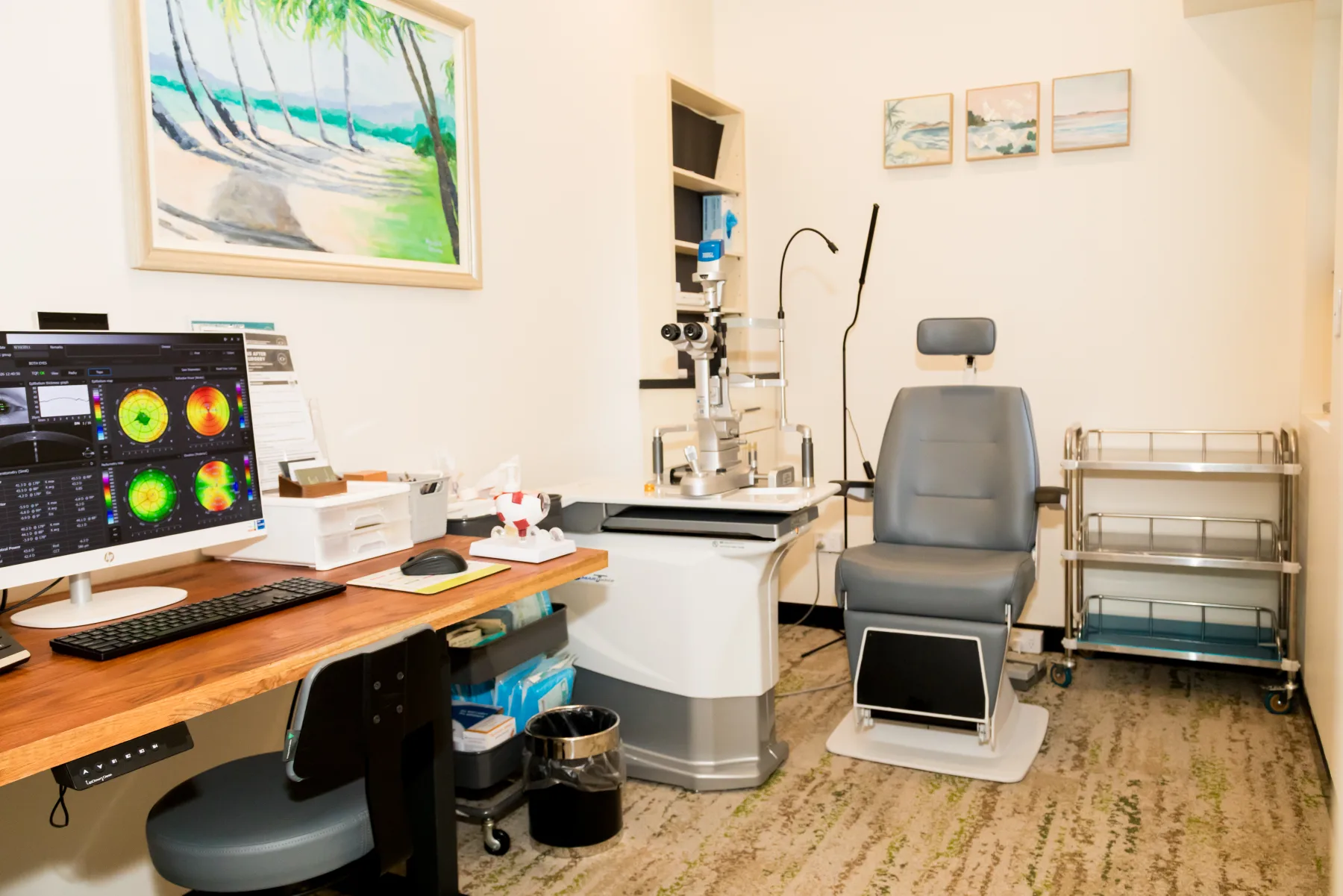
Every examination and treatment plan is shaped around your symptoms, test results and individual needs. The equipment shown is part of our real clinical environment; the tests you need are selected after assessment.
See inside the practice ↗Symptoms vary between people, and some conditions cause no early warning. A specialist examination is needed for an accurate diagnosis.
Vitrectomy is recommended for a diagnosed condition rather than one symptom alone
Persistent blurred or distorted vision from vitreoretinal disease
Vision loss from non-clearing vitreous haemorrhage
Symptoms associated with a retinal detachment or macular disorder
A visual summary of the condition, including common symptoms, risk factors, assessment and available treatment approaches. This guide supports—but does not replace—a specialist examination.
View full-size guide ↗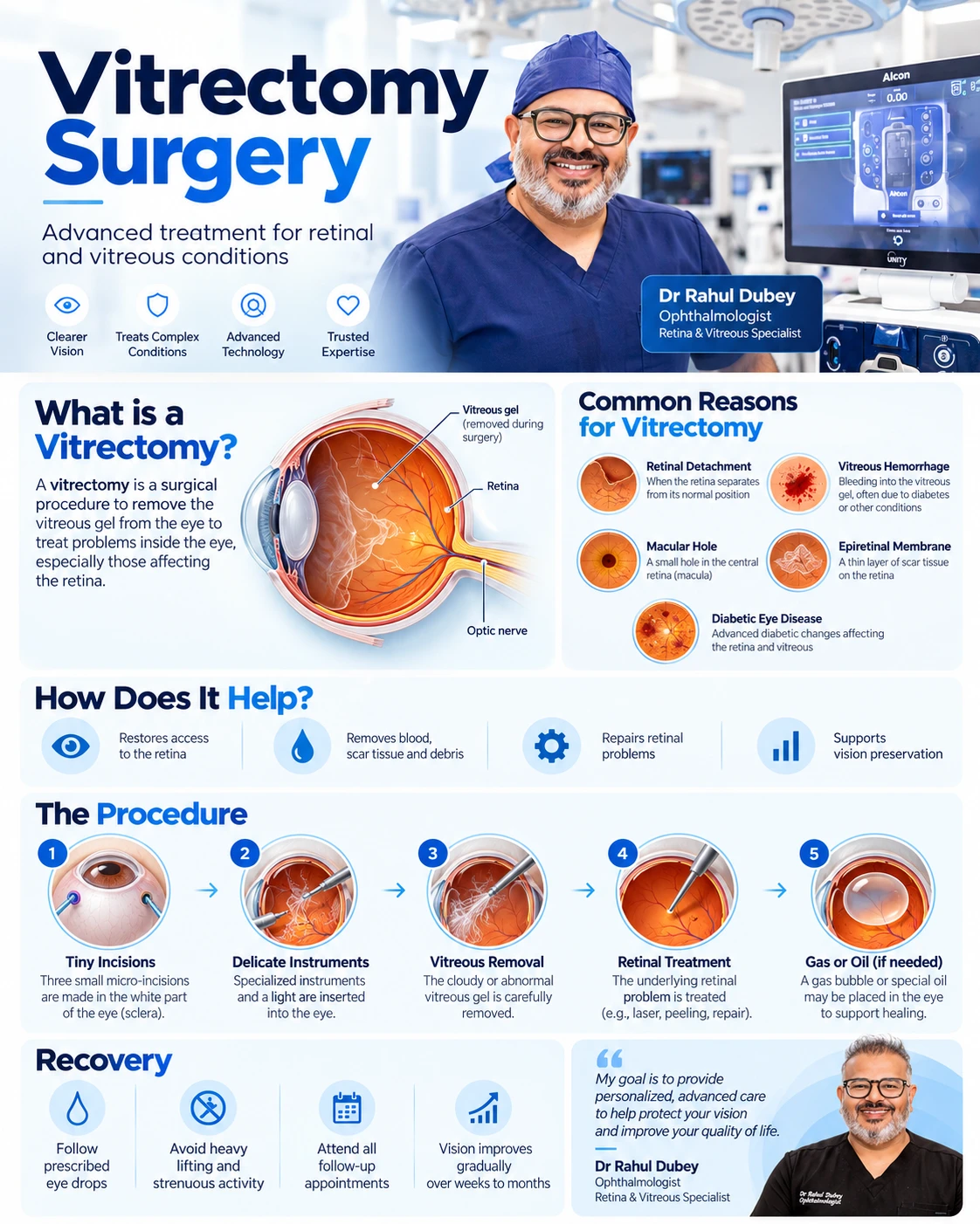
Having a risk factor does not confirm the condition. Your history and examination are considered together.
Examination and imaging establish whether surgery is likely to offer a meaningful anatomical or visual benefit.
High-resolution imaging helps define macular membranes, holes, swelling and other retinal features.
The procedure, anaesthesia, possible gas or oil, positioning and realistic outcomes are discussed before consent.
Findings are explained in context so the recommended next step reflects your eyes, health and visual needs.
Use prescribed drops and eye protection exactly as directed.
Follow any face-positioning instructions carefully if a gas bubble or oil is used.
Do not fly or travel to high altitude while an intraocular gas bubble remains; confirm restrictions with your surgeon.
Attend all scheduled reviews and report increasing pain, redness or worsening vision promptly.
These answers are general. Your own care plan may differ after examination.
It is used for selected problems such as retinal detachment, macular hole, epiretinal membrane, vitreous haemorrhage and other vitreoretinal conditions.
Not in every operation. The need for gas, air or silicone oil depends on the condition and surgical plan.
Recovery varies with the condition, procedure and starting health of the retina. Your surgeon will explain a realistic expected range.
Yes. The recommendations, timing and follow-up are based on your examination, imaging, general health and visual priorities.