Dilated retinal examination
The pattern of bleeding and vessel change identifies the type of occlusion.
Treatment and monitoring for blocked retinal veins.
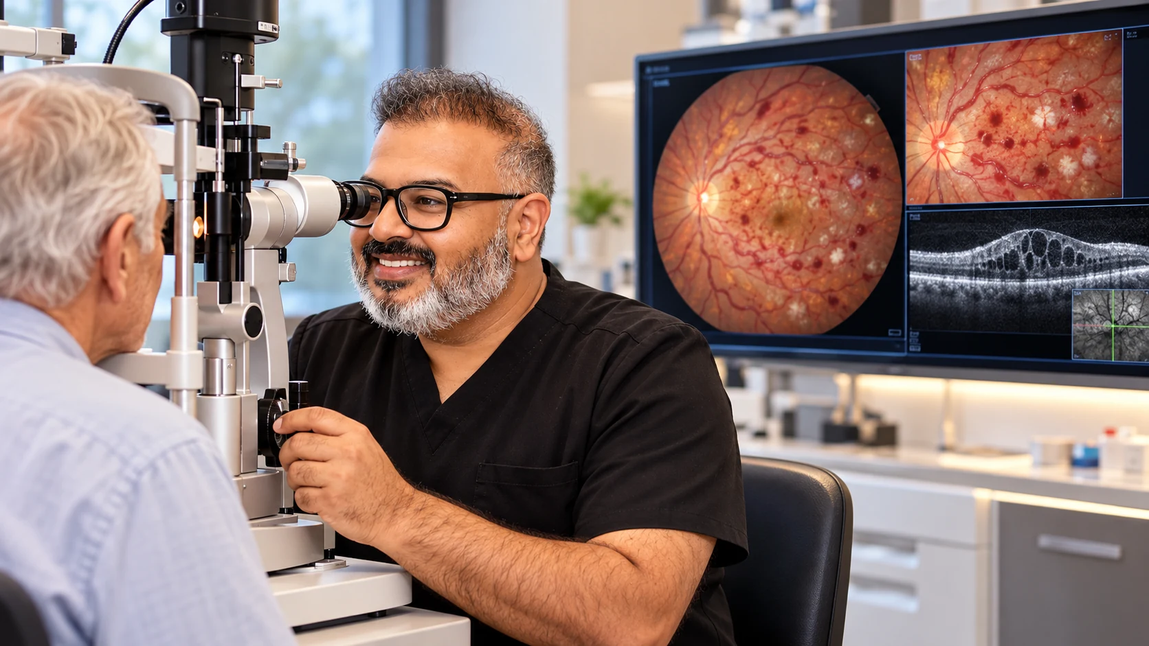
A retinal vein occlusion can cause bleeding, swelling and painless blurred vision.
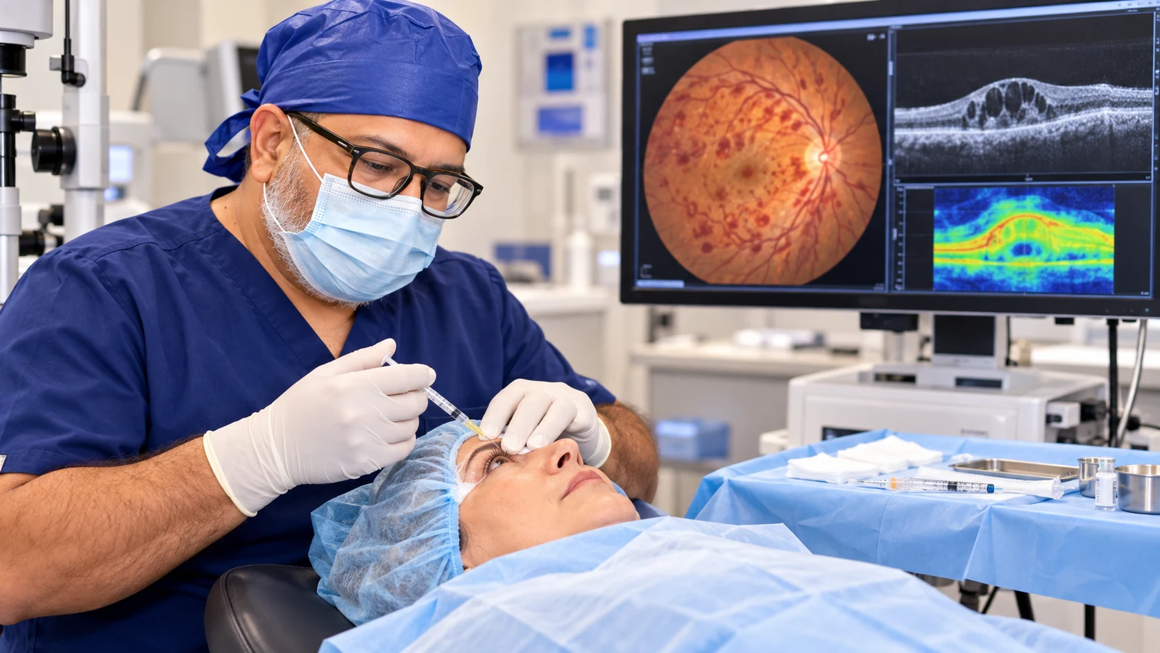
Every examination and treatment plan is shaped around your symptoms, test results and individual needs. The equipment shown is part of our real clinical environment; the tests you need are selected after assessment.
See inside the practice ↗Symptoms vary between people, and some conditions cause no early warning. A specialist examination is needed for an accurate diagnosis.
Sudden or gradual painless blur in one eye
Distorted central vision
A dark or missing patch
Floaters caused by retinal bleeding
Reduced contrast or difficulty reading
A visual summary of the condition, including common symptoms, risk factors, assessment and available treatment approaches. This guide supports—but does not replace—a specialist examination.
View full-size guide ↗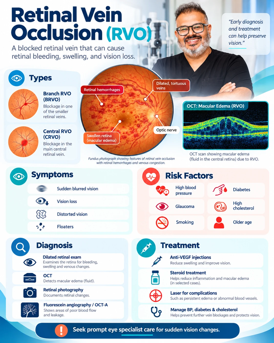
Having a risk factor does not confirm the condition. Your history and examination are considered together.
The pattern of bleeding and vessel change identifies the type of occlusion.
Imaging measures macular swelling and guides treatment.
Dye imaging may map circulation, leakage and areas of poor perfusion.
Blood pressure and vascular risk factors are reviewed with the patient’s medical team.
Attend OCT monitoring because swelling can recur without an obvious early symptom.
Keep injection or laser appointments on the recommended schedule.
Work with your GP to address blood pressure and other vascular risks.
Report pain, redness, sudden worsening or new floaters promptly.
These answers are general. Your own care plan may differ after examination.
It is a blockage in a retinal vein. It prompts review of vascular health but is not the same event as a brain stroke.
Recovery varies with the vessel involved, macular damage, circulation and response to treatment.
The course is guided by OCT and disease activity; some people require ongoing treatment.
Risk depends on general health and eye factors, so managing these and monitoring both eyes are important.