Vision and distortion testing
Vision is measured and each eye is assessed for central distortion.
Assessment and microsurgical care for a fine membrane over the macula.

An epiretinal membrane can wrinkle the retinal surface and distort central vision.
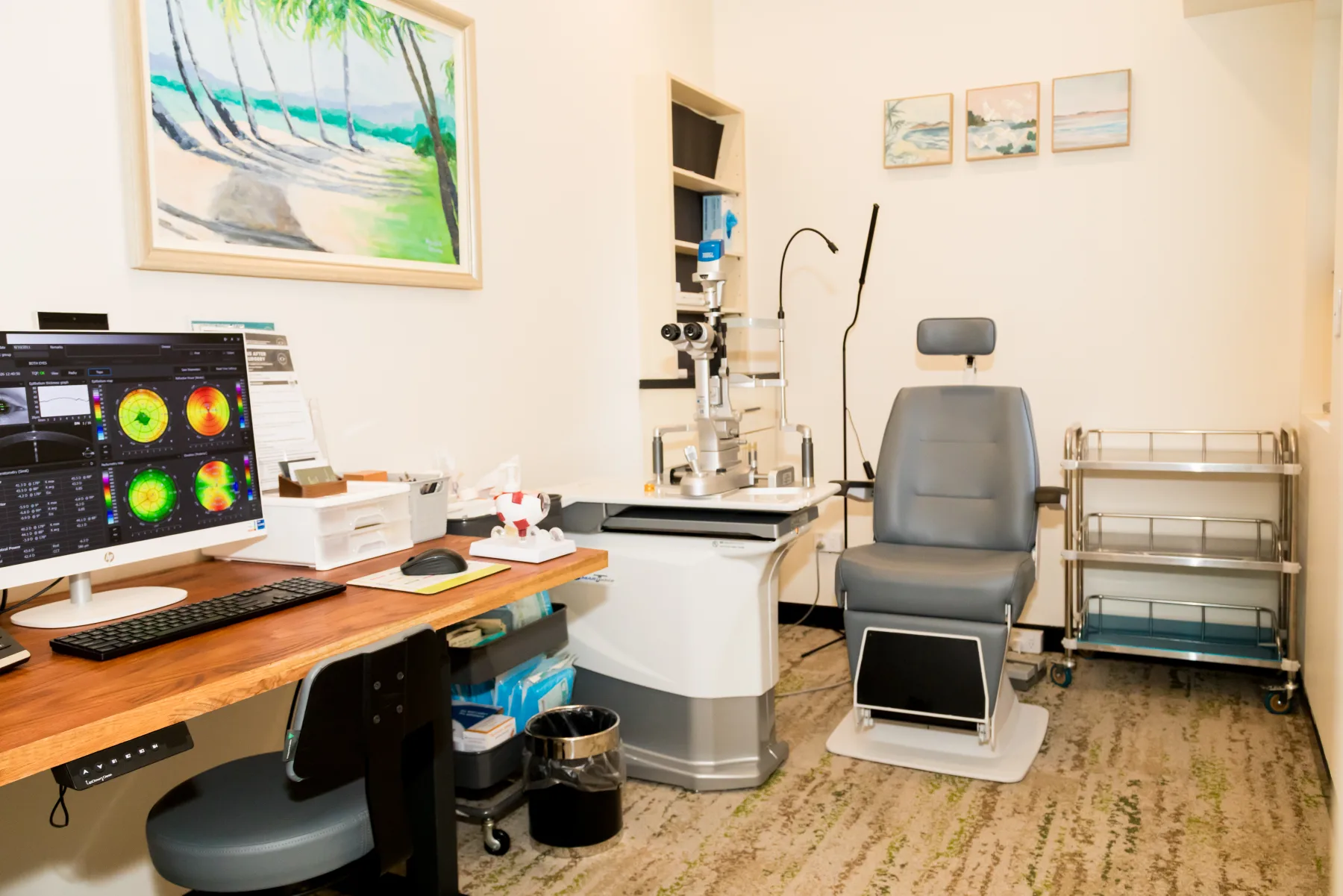
Every examination and treatment plan is shaped around your symptoms, test results and individual needs. The equipment shown is part of our real clinical environment; the tests you need are selected after assessment.
See inside the practice ↗Symptoms vary between people, and some conditions cause no early warning. A specialist examination is needed for an accurate diagnosis.
Straight lines appearing bent or wrinkled
Blurred central vision
Difficulty reading fine print
Images appearing different in size between the eyes
Double or ghosted vision in one eye
Having a risk factor does not confirm the condition. Your history and examination are considered together.
Vision is measured and each eye is assessed for central distortion.
The retinal surface is examined for membrane, traction and related disease.
Cross-sectional images show the membrane and its effect on the macular layers.
Previous scans and symptoms help determine whether the condition is stable or progressing.
Use prescribed drops and attend postoperative reviews.
Blur and mild discomfort can occur early; improvement is usually gradual.
Follow advice about activity, eye protection and any gas-bubble restrictions.
Report increasing pain, redness or sudden worsening of vision promptly.
These answers are general. Your own care plan may differ after examination.
No. Surgery is considered when symptoms and OCT findings justify the potential benefit and risk.
It often improves, but the final result depends on duration, severity and retinal health.
Recurrence is uncommon but possible, so follow-up remains important.
Vitrectomy can accelerate cataract in an eye with its natural lens; this is discussed during planning.