Dilated macular examination
The retina is checked for fluid and to exclude other macular disorders.
Monitoring and treatment for fluid beneath the central retina.

Fluid beneath the macula can cause blur, distortion or altered image size.
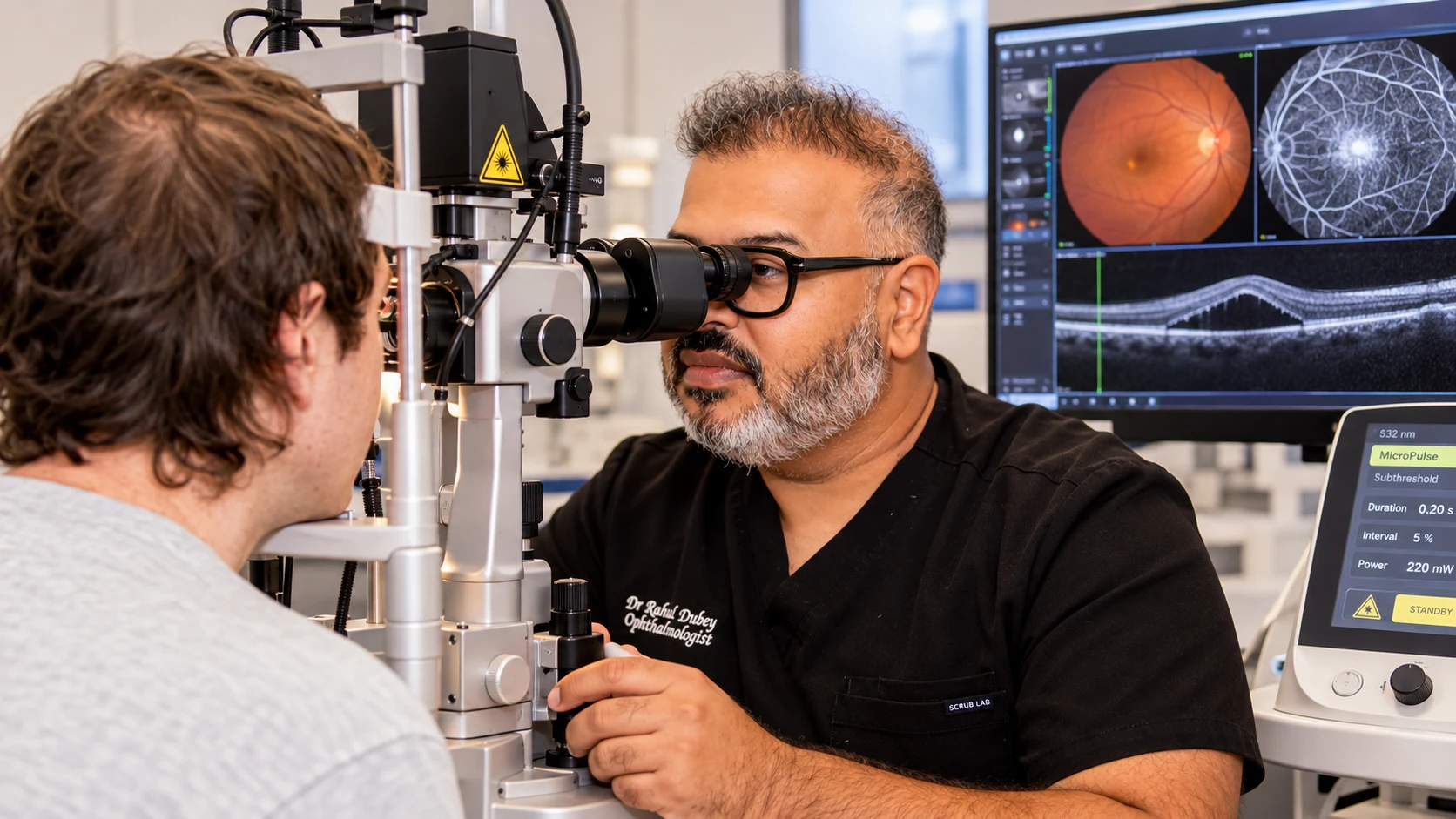
Every examination and treatment plan is shaped around your symptoms, test results and individual needs. The equipment shown is part of our real clinical environment; the tests you need are selected after assessment.
See inside the practice ↗Central serous retinopathy can blur or distort the centre of your vision. This visual comparison is an illustration only—not a promise of a particular treatment result. Recovery and treatment needs vary between patients and are discussed after specialist assessment.

Symptoms vary between people, and some conditions cause no early warning. A specialist examination is needed for an accurate diagnosis.
Blurred or dim central vision in one eye
Straight lines appearing bent
Objects appearing smaller or farther away
Reduced contrast or altered colour
A central grey or dark patch
A visual summary of the condition, including common symptoms, risk factors, assessment and available treatment approaches. This guide supports—but does not replace—a specialist examination.
View full-size guide ↗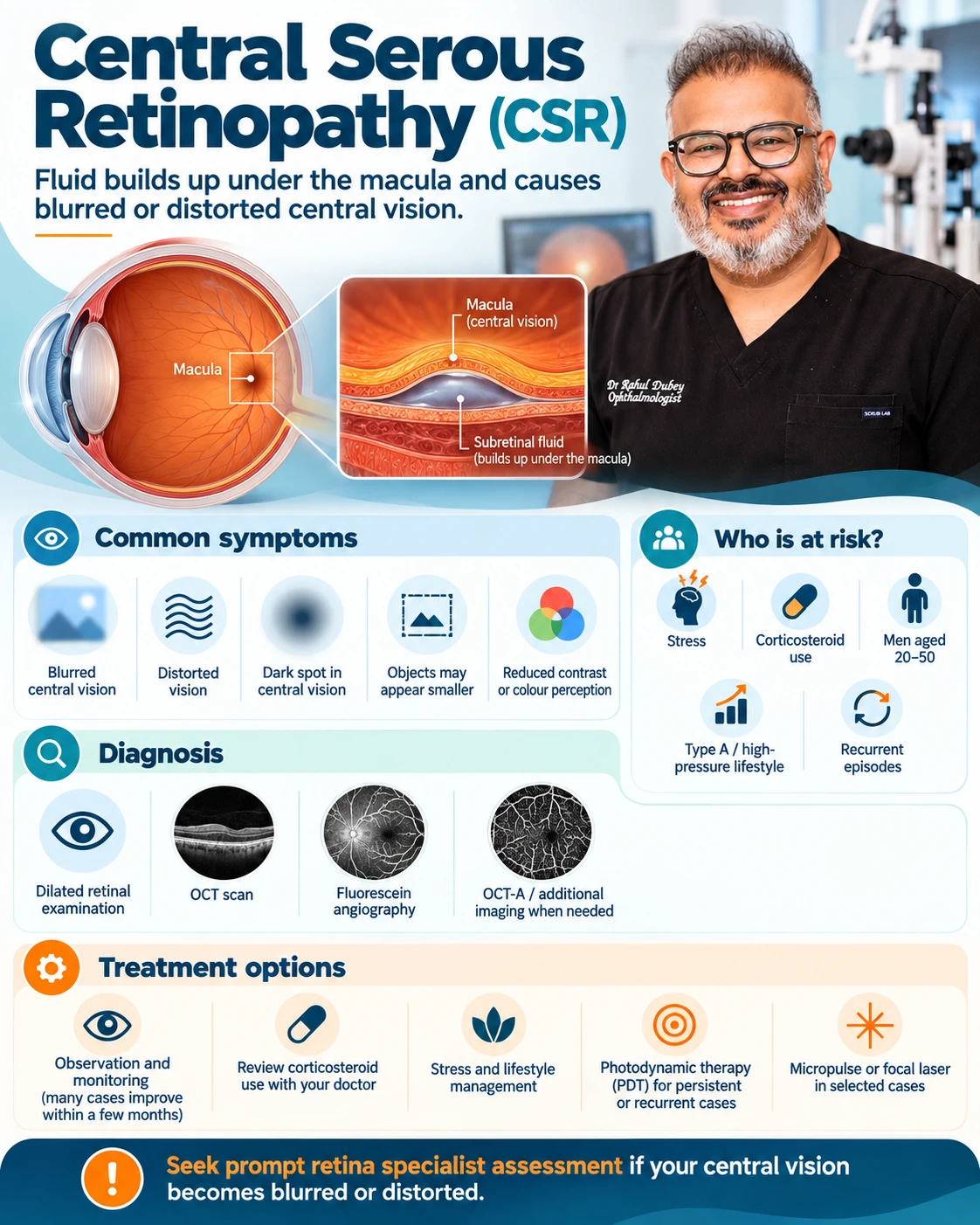
Having a risk factor does not confirm the condition. Your history and examination are considered together.
The retina is checked for fluid and to exclude other macular disorders.
Cross-sectional imaging confirms and measures fluid beneath the retina.
This may reveal patterns of retinal pigment epithelium stress or previous episodes.
Fluorescein or indocyanine green angiography may locate leakage in persistent or atypical disease.
Attend OCT reviews until fluid and vision stabilise.
Report recurrence of distortion or central blur.
Discuss steroid exposure with the prescribing doctor rather than stopping it independently.
Address sleep apnoea, stress and general health factors where relevant.
These answers are general. Your own care plan may differ after examination.
Many first episodes resolve, but persistent or recurrent fluid can affect retinal function and needs monitoring.
Stress is associated with it, but the condition is multifactorial and should not be attributed to stress alone.
Do not stop prescribed steroids suddenly. Discuss the risk and possible alternatives with the prescribing doctor and eye specialist.
It can reduce leakage in selected persistent or recurrent cases after imaging identifies the affected area.