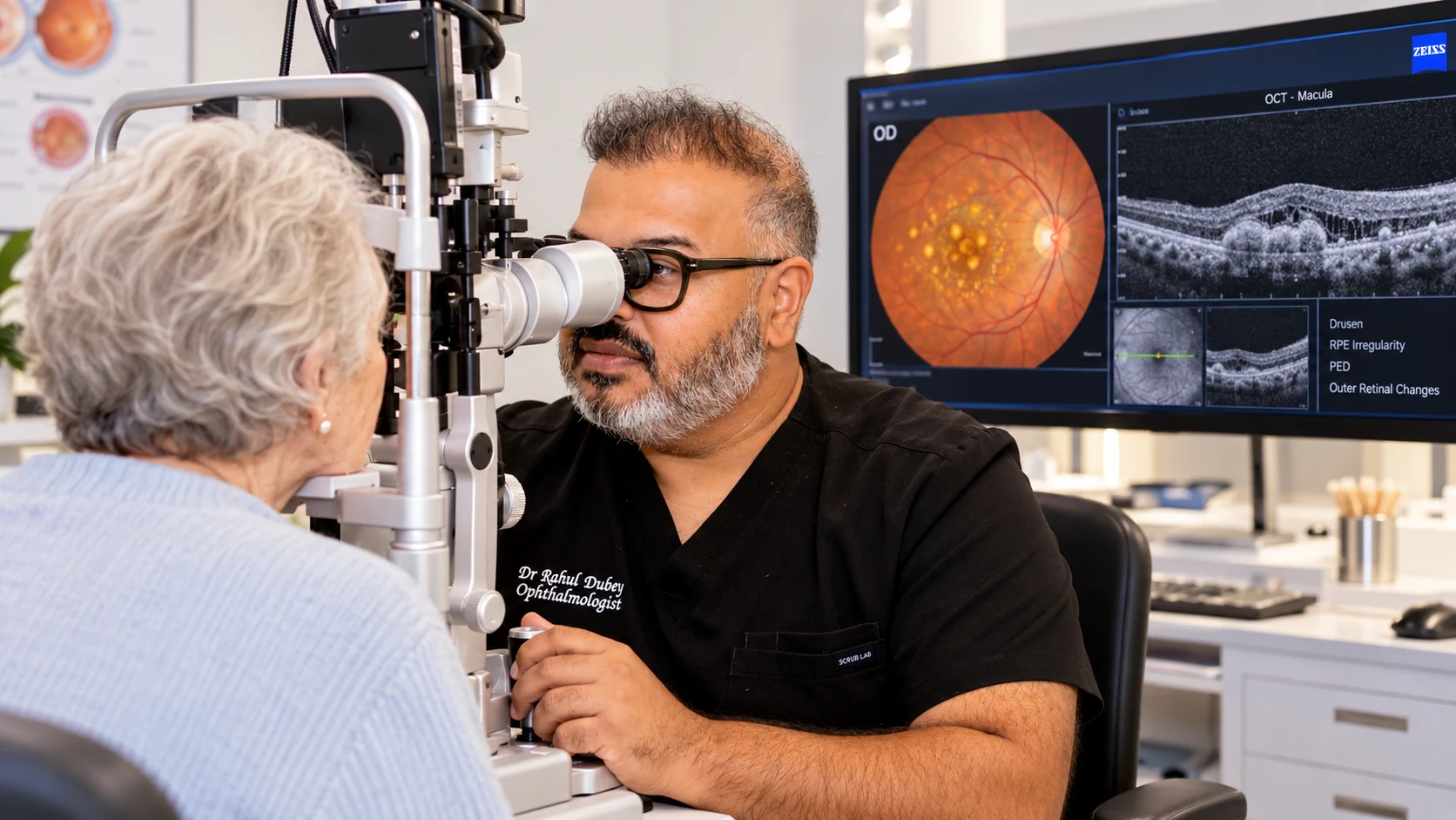
Dilated macular examination
The macula is examined for drusen, pigment change, bleeding or fluid.
Monitoring and treatment for dry and wet AMD.
AMD affects the macula, the central area responsible for detailed vision.
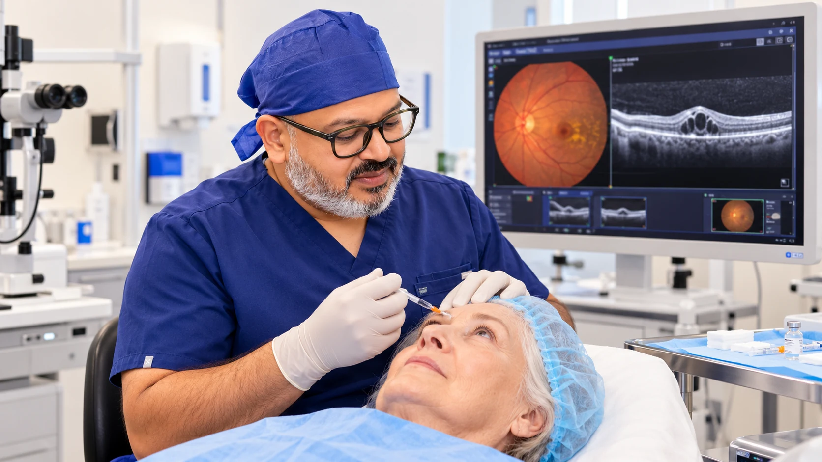
Every examination and treatment plan is shaped around your symptoms, test results and individual needs. The equipment shown is part of our real clinical environment; the tests you need are selected after assessment.
See inside the practice ↗Symptoms vary between people, and some conditions cause no early warning. A specialist examination is needed for an accurate diagnosis.
Straight lines appearing wavy or bent
Blurred or missing central vision
Difficulty reading or recognising faces
Reduced contrast or need for brighter light
A new central grey or dark patch
Having a risk factor does not confirm the condition. Your history and examination are considered together.
The macula is examined for drusen, pigment change, bleeding or fluid.
High-resolution imaging detects fluid and maps retinal layers.
Photographs document appearance and support comparison over time.
Dye or OCT angiography may help identify abnormal vessels when wet AMD is suspected.
Monitor each eye separately as instructed and report new distortion promptly.
Attend injection and OCT appointments on the recommended schedule.
After injection, mild grittiness can occur; increasing pain or redness needs urgent advice.
Continue general-health measures and use prescribed supplements only after professional advice.
These answers are general. Your own care plan may differ after examination.
AMD affects central detail rather than usually removing all peripheral vision, but it can significantly affect daily activities.
Yes. A new rapid visual change or distortion should be assessed promptly.
They control abnormal vessel activity; repeated treatment and monitoring are often required.
No. AREDS2-type supplements are recommended only for selected stages and should be discussed with an eye-care professional.